Introduction
Chapman points are small, palpable, tender nodules located in specific anatomic regions, representing reflexes linked to visceral dysfunction. These points may be helpful in osteopathic manipulative treatment (OMT) as diagnostic and therapeutic indicators. Understanding and effectively addressing Chapman points can support visceral diagnoses. This article details the anatomic distribution, diagnostic significance, and therapeutic approaches to Chapman points throughout the body.
Pathophysiology
Chapman points are characteristically discrete, firm nodules approximately 2-3 mm in diameter. Typically located within the deep fascial layers, periosteum of bones, tendinous attachments, or subcutaneous tissue, these points reflect viscerosomatic reflexes mediated primarily by the sympathetic nervous system. Dysfunction or pathology in a visceral organ leads to localized lymphatic congestion and increased fascial tension, manifesting as exquisitely tender nodules in corresponding anatomic areas.
Location of Chapman points
In general, each organ has an anterior
The following sections list the Chapman points for various regions of the body. Minor variations exist between sources, occasionally differing by one segmental level (eg, T1 vs T2). For clinical and examination purposes, such precise differentiation is generally not required

Head and neck
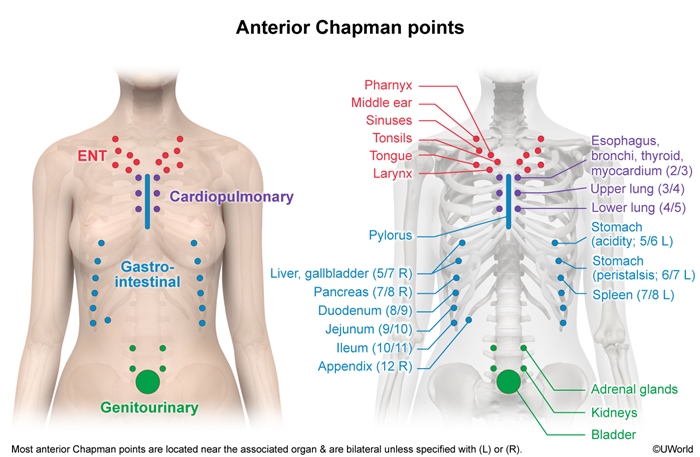
Sinuses
- Anterior: Midclavicle.
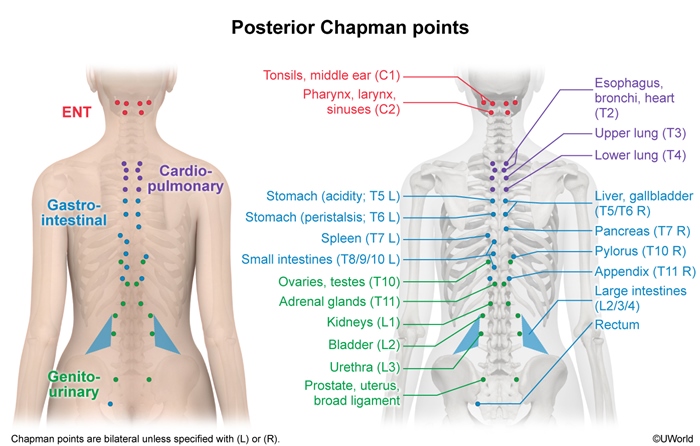
- Posterior: C2.
Tonsils
- Anterior: Between ribs 1 and 2 near sternum.
- Posterior: C1 posterior arch.
Middle ear
- Anterior: Superior clavicle at crossing of first rib.
- Posterior: C1.
Thorax
Esophagus/thyroid/bronchi
- Anterior: Second to third intercostal space (ICS) near sternum.
- Posterior: T2.
Heart
- Anterior: Second to third ICS near sternum (left side).
- Posterior: Between T2 and T3 (left transverse processes [TPs]).
Bronchi
- Anterior: Second ICS near sternum.
- Posterior: T2.
Lungs
- Anterior: Third to fourth ICS (upper lungs) and fourth to fifth ICS near sternum.
- Posterior: T3-T4 (upper) and T4-T5 (lower).
Gastrointestinal tract
Stomach
- Anterior: Fifth to seventh ICS on left.
- Posterior: T5-T7 on left.
Liver and gallbladder
- Anterior: Fifth to seventh ICS on right.
- Posterior: T5-T7 on right.
Spleen
- Anterior: Ribs 7-8 (left) at costochondral junction (CCJ).
- Posterior: T7-T8 on left.
Pancreas
- Anterior: Ribs 7-8 (right) at CCJ.
- Posterior: T7-T8 on right.
Small intestine
- Anterior: Near the CCJ, ribs 8-9 (duodenum), ribs 9-10 (jejunum), ribs 10-11 (ileum).
- Posterior: T8-T9 (duodenum), T9-T10 (jejunum), T10-T11 (ileum).
Appendix
- Anterior: Tip of the right twelfth rib.
- Posterior: Right T11-T12 TP.
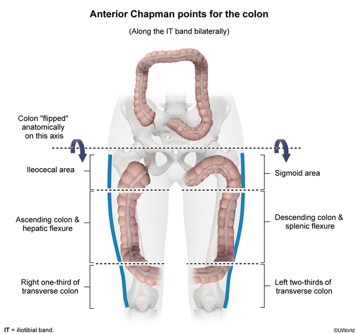
Colon
- Anterior: Iliotibial bands (mapped along pattern of colon).
- Posterior: L2-L4 TP.
Genitourinary
Kidneys
- Anterior: 1 inch lateral and 1 inch superior to umbilicus.
- Posterior: T12-L1.
Adrenal glands
- Anterior: 2 inches superior and 1 inch lateral to umbilicus.
- Posterior: T11-T12.
Bladder
- Anterior: Periumbilical region.
- Posterior: L2.
Urethra
- Anterior: Pubic symphysis.
- Posterior: L3.
Prostate
- Anterior: Lateral femur along the posterior iliotibial band.
- Posterior: Between L5 TP and posterior superior iliac spine.
Ovaries/testes
- Anterior: Pubic symphysis/anterior pubic bone.
- Posterior: T9-T11.
Uterus
- Anterior: Medial edge of obturator foramen.
- Posterior: L5.
Therapeutic applications
Integrating Chapman points into clinical practice enhances diagnostic clarity and therapeutic precision. Using these points as part of a comprehensive osteopathic examination facilitates early detection and management of visceral dysfunction. Effective management of Chapman points involves a gentle, rotary stimulation technique directly applied to the identified nodule. This treatment aims to:
- Alleviate tenderness.
- Decrease lymphatic congestion.
- Normalize sympathetic nervous system function.
The therapeutic steps include:
- Precise palpation and identification of the Chapman point.
- Application of gentle rotary pressure using fingertip pads for 10-30 seconds.
- Reassessment posttreatment for reduced tenderness and decreased nodule size.
Chapman point therapy synergistically complements other OMT techniques, including myofascial release, lymphatic pump techniques, and muscle energy treatments.
Summary
Chapman points are small, palpable nodules associated with viscerosomatic reflexes that serve both diagnostic and therapeutic purposes in osteopathic manipulative treatment. Located in predictable anterior and posterior anatomic regions, these 2- to 3-mm, tender nodules reflect dysfunction in visceral organs through the sympathetic nervous system, often due to lymphatic congestion and fascial tension. Their precise locations correspond to various systems—including respiratory, gastrointestinal, and genitourinary—enhancing diagnostic accuracy for visceral dysfunction. In addition to treating the associated underlying medical condition, Chapman points are managed using gentle rotary pressure, which helps reduce tenderness, restore autonomic balance, and improve lymphatic flow.